Adult Hip Dysplasia
What is Adult Hip Dysplasia?
Adult hip dysplasia is a condition in which the hip joint does not develop normally during childhood and adolescence and continues to affect the hip in adulthood. It is one of the more common conditions Dr Williams sees in young female patients with hip pain.
The hip is usually a highly stable ball-and-socket joint. In hip dysplasia, the socket (acetabulum) is too shallow or poorly shaped, meaning it does not fully cover or support the ball (femoral head). This places abnormal stress on the cartilage, labrum, and surrounding structures. As with most conditions, there is a broad spectrum of severity, ranging from a mildly shallow socket in a well-functioning hip to a completely dislocated hip.
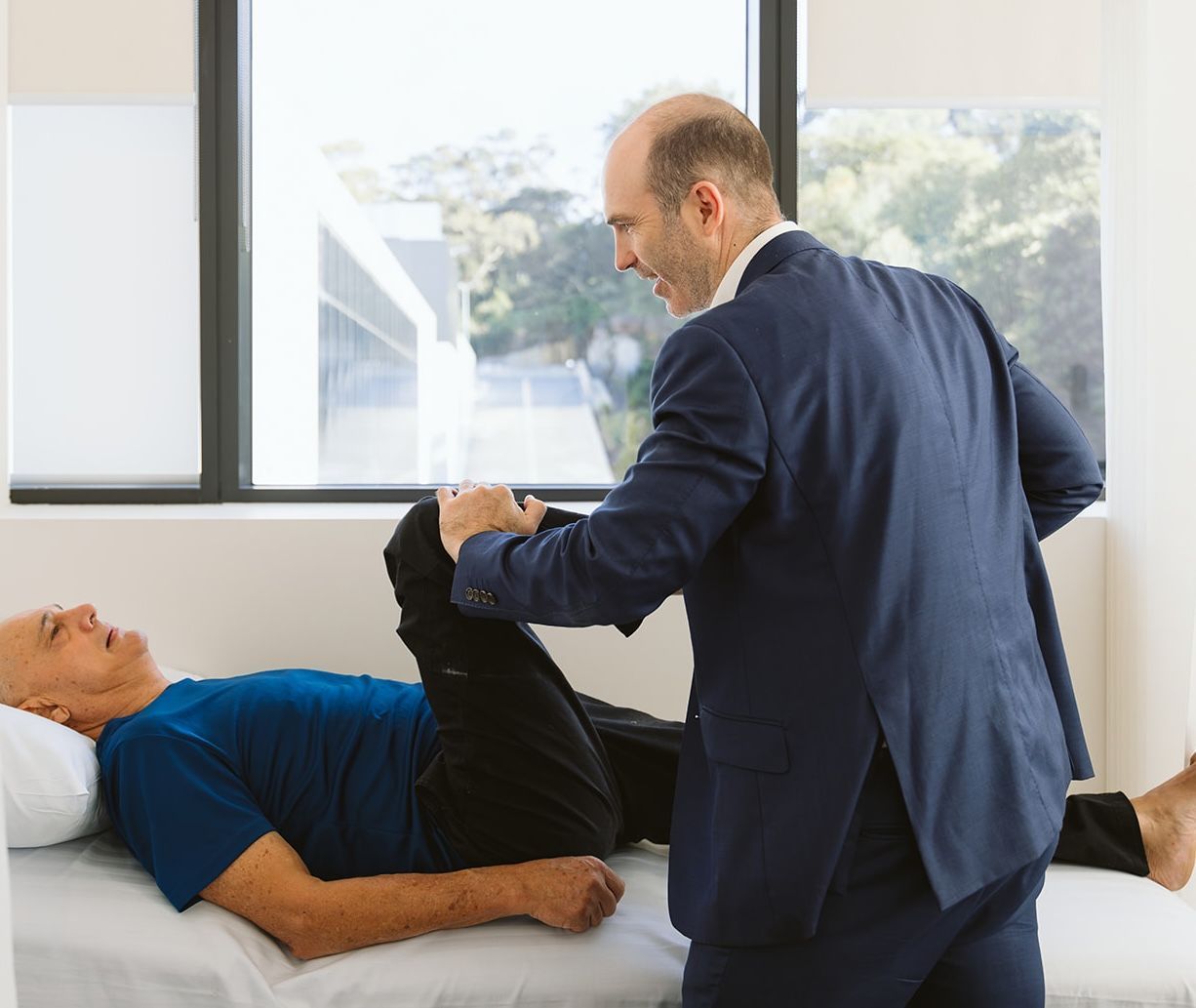
While hip dysplasia is often thought of as a condition diagnosed in infancy or childhood, many individuals are not diagnosed until adolescence or adulthood. This is because most forms of mild and moderate hip dysplasia are very hard to detect with clinical examination, and Australia has a selective ultrasound screening program, meaning only babies with risk factors or clinical concerns generally have a scan.
Many adults may have mild dysplasia and remain symptom-free for years, while others develop pain, stiffness, or early osteoarthritis. Adult hip dysplasia is a recognised cause of hip pain in young and middle-aged adults and is a leading cause of early hip arthritis.
The condition can affect one or both hips.
How Does Adult Hip Dysplasia Impact Your Hip and Health?
Adult hip dysplasia alters the normal mechanics of the hip joint. In a healthy hip, the socket distributes forces evenly across the joint surface during walking, standing, and other movements. In dysplasia, the shallow socket concentrates stress on a smaller area of cartilage and bone.
Key anatomical and health impacts include:
- Cartilage wear: Increased pressure and sheer forces accelerate cartilage breakdown, leading to early osteoarthritis
- Labral damage: The labrum, a ring of cartilage that stabilises the hip, is prone to overload and tearing due to joint instability
- Joint instability: Poor coverage of the femoral head allows excessive movement within the joint
- Muscle fatigue: Surrounding muscles work harder to stabilise the hip, leading to fatigue and overuse pain
- Altered posture and gait: Compensation may affect the lower back, pelvis, knees, and opposite hip
Over time, these changes can lead to chronic pain, reduced mobility, and difficulty with activities of daily living, such as walking, standing, sitting, and exercising. If untreated, adult hip dysplasia can increase the risk of developing hip osteoarthritis at a younger age than expected.
Causes and Risk Factors for Adult Hip Dysplasia
Certain individuals are at increased risk of developing or being diagnosed with adult hip dysplasia. Risk factors include:
- Women: Adult hip dysplasia is more common in women than in men
- History of childhood hip problems: Including developmental dysplasia of the hip (DDH) that was untreated or partially treated
- Family history: Genetic factors increase the likelihood of hip dysplasia
- First-born children: Associated with higher rates of childhood hip dysplasia
- Breech birth: Increases the risk of abnormal hip development
- Joint hypermobility: Increased ligament laxity can worsen hip instability
Many adults with hip dysplasia may not realise they have a structural hip issue until symptoms appear, often in their 20s to 40s.
Symptoms of Adult Hip Dysplasia
Symptoms of adult hip dysplasia vary with the severity of the condition and the extent of joint damage. Common symptoms include:
- Hip or groin pain: Often felt at the front of the hip, deep in the groin or deep in the side/back of the hip.
- Pain with activity: Especially walking, running, climbing stairs, or prolonged standing
- Hip stiffness: Reduced range of motion may be a symptom once arthritic change is established. In the early stages, the hip range may actually be increased compared to the average due to the shallow socket
- Clicking, catching, locking or feeling of instability: Often related to labral tears
- Pain when sitting: Discomfort during long periods of sitting or driving
- Limping: Changes in walking pattern due to pain or instability
- Lower back or buttock pain: Caused by altered movement mechanics
Symptoms may start gradually and worsen over time. Some people experience flare-ups, while others develop constant pain that interferes with work, exercise, and daily activities.
Preventing Adult Hip Dysplasia
True prevention of adult hip dysplasia is limited because the condition develops early in life. If you have been diagnosed with adult hip dysplasia, it is worth remembering to screen any future children by requesting your GP to order an ultrasound at 6 weeks of age. In diagnosed adults, progression of symptoms and joint damage can often be slowed with appropriate strategies.
Preventive and protective measures include:
- Activity modification: Avoiding repetitive high-impact activities that overload the hip joint
- Maintaining a healthy weight: Reduces stress on the hip joint
- Strengthening exercises: Targeting hip, core, and pelvic muscles to improve joint support. As the bone support is insufficient, strengthening the dynamic stabilisers of the hip is extremely important.
- Prompt treatment of symptoms: Early assessment when hip pain begins can prevent further damage
- Avoiding prolonged positions: Limiting long periods of sitting or standing without movement
For some individuals, non-surgical management such as physiotherapy, pain management strategies, and activity modification can effectively control symptoms. In others, especially younger adults with significant dysplasia and preserved cartilage, early surgical intervention may be recommended to preserve the joint and delay or prevent arthritis.
Progression of Adult Hip Dysplasia
Adult hip dysplasia tends to progress over time, particularly if joint instability is not addressed. The stages are usually defined by symptoms, joint mechanics, and cartilage health rather than age alone.
- Structural abnormality without symptoms: The hip socket is shallow, but cartilage and labrum remain relatively healthy. Many people are unaware they have dysplasia
- Early symptoms and labral strain: Intermittent hip or groin pain appears, often during activity. Labral tears may develop
- Progressive cartilage damage: Pain becomes more frequent, stiffness increases, and imaging shows cartilage thinning and joint overload
- Early osteoarthritis: Joint space narrowing, bone spurs, and reduced hip motion are present
- Advanced osteoarthritis: Severe pain, marked stiffness, loss of function, and structural joint collapse
Progression is influenced by the severity of dysplasia, activity level, body weight, muscle strength, and the timing of diagnosis and management.
Diagnosis of Adult Hip Dysplasia
Diagnosing adult hip dysplasia requires a combination of clinical assessment and careful evaluation of imaging. Because symptoms can mimic other more common hip conditions, such as femoroacetabular impingement, a diagnosis of hip dysplasia can often be overlooked or delayed.
Clinical assessment includes:
- Detailed medical history: Onset, location, duration, and triggers of hip pain (particularly prolonged standing/walking/running)
- Physical examination: Hip range of motion (often excessive), strength, stability (often poor control of single-leg squat), and gait assessment (occasional limp, intoeing gait)
Imaging studies commonly include:
- X-rays: The primary diagnostic tool. Routine assessment includes a standing pelvic x-ray as well as a ‘false profile’ view of the affected hip. The centre-edge angle is the primary diagnostic measurement of dysplasia. The normal centre edge angle is 25-40 degrees. 18-25 degrees is considered borderline dysplasia, and less than 18 degrees is considered true dysplasia.
- Secondary diagnostic measures include: the Tonnis angle, which measures the upslope of the acetabular roof (normal 0-10 degrees); the anterior centre edge angle (ACEA) seen on the false profile view; and a reduced anterior wall index.
- MRI or MR arthrogram: Evaluates labral tears, cartilage damage, and soft tissue structures
- CT scan: May be used to measure the rotational profile of the lower limbs and 3D anatomy of the hip socket if surgery is being considered.
Early diagnosis is important, as treatment options are broader before significant cartilage damage occurs.
Treatment for Adult Hip Dysplasia
Treatment depends on age, symptom severity, activity level, and cartilage condition. Dr Williams usually recommends a stepwise approach.
Non-surgical Treatment Options
These are often recommended for mild to moderate symptoms or early-stage disease:
- Activity modification: Reducing high-impact or repetitive hip-loading activities
- Physiotherapy: Strengthening hip stabilisers, core muscles, and improving movement patterns. Dedication to a supervised strengthening program is crucial for success.
- Shoe raise: In select cases of ‘functional dysplasia’ contributed to by a leg length difference, a shoe raise can be helpful
- Pain management: Anti-inflammatory medications or targeted injections where appropriate
- Weight management: Reducing load across the hip joint
- Lifestyle adjustments: Limiting prolonged sitting, standing, or uneven terrain
Non-surgical care aims to control symptoms but does not correct the underlying bone structure.
Surgical Treatment Options for Adult Hip Dysplasia
Dr Williams may recommend surgery when non-operative options have not led to adequate symptom control. The two main types of surgery that Dr Williams performs are joint-preserving surgery or joint-replacing surgery.
- Periacetabular osteotomy (PAO): For patients between 13 and 40 with minimal arthritis, a PAO can reorient the hip socket to improve coverage and stability, and prevent or delay the need for hip replacement.
- Hip arthroscopy: May be used to treat labral tears or cartilage damage, but is usually combined with a PAO in dysplastic hips. Hip arthroscopy alone is accepted to have poorer outcomes in dysplastic hips and can even worsen symptoms.
- Femoral osteotomy: In patients with acetabular dysplasia, there is often a degree of femoral dysplasia, most commonly in the form of increased femoral anteversion. In severe cases, a femoral derotation osteotomy may be required in addition to PAO to restore hip biomechanics.
- Total hip replacement: If the dysplasia has led to significant arthritis or the patient is over 40, a total hip replacement may be a more reliable surgical option for treating dysplasia-related pain.
Early intervention with joint-preserving surgery, when appropriate, can significantly delay or prevent the need for hip replacement. It is a major decision, however, to undergo periacetabular osteotomy, and Dr Williams will carefully discuss the risks and benefits and whether it is appropriately indicated in your particular case.
What if Adult Hip Dysplasia is Untreated?
Leaving adult hip dysplasia untreated can lead to predictable and progressive joint deterioration over time. The speed of progression generally depends on the severity of the dysplasia, and patients with milder cases may do well until needing a hip replacement in their 60’s or 70’s, which is a perfectly acceptable outcome.
Untreated dysplasia imposes abnormal forces on the joint with every hip load. Over the years, this mechanical overload eventually leads to irreversible cartilage breakdown. Adult hip dysplasia is one of the most common reasons Dr Williams needs to perform total hip replacements in adults under 50.
One of the risks of taking a ‘wait and see’ approach in a young person with symptomatic hip dysplasia is that joint degeneration may progress to a point that hip preservation surgery is no longer appropriate.