Baby Hip Dysplasia (DDH)
What is Baby Hip Dysplasia (DDH)?
Baby Hip Dysplasia, medically known as Developmental Dysplasia of the Hip (DDH), is a condition where a baby’s hip joint does not form properly. The hip is a ball-and-socket joint. In DDH, the socket (acetabulum) may be too shallow, allowing the ball (the head of the thigh bone or femur) to move out of place. This can range from mild looseness of the joint to a complete dislocation.
DDH can be present at birth or develop during the first months of life. Some babies are born with unstable hips that may later become normal, while others develop increasing instability if the condition is not recognised early. The condition affects one or both hips, with the left hip being more commonly involved. DDH is relatively common, particularly in newborns, and early detection is important because treatment is usually simpler and more effective when started early.
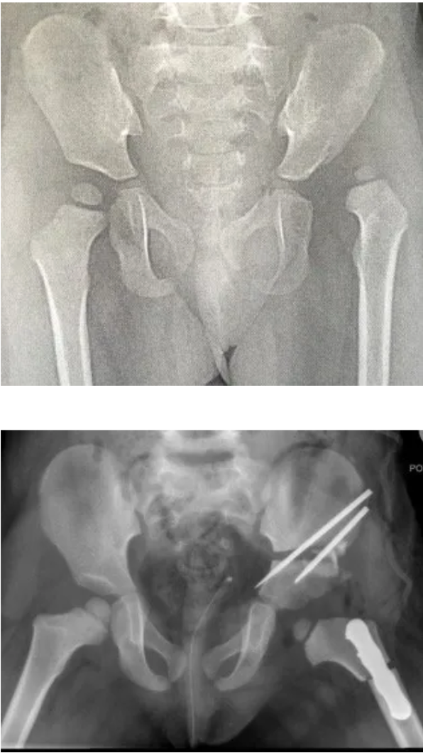
How Does Baby Hip Dysplasia (DDH) Impact Your Child’s Hip?
In a healthy hip, the rounded femoral head sits securely within the hip socket, allowing smooth movement and even distribution of forces as the baby grows and later starts to crawl, stand, and walk.
In DDH, this normal relationship is altered.
- Shallow hip socket: The socket may not be deep enough to fully cover the femoral head, reducing stability.
- Loose ligaments: The soft tissues around the hip may be more flexible than normal, allowing excessive movement.
- Partial or full dislocation: In more severe cases, the femoral head may sit partly or completely outside the socket.
When the ball is poorly positioned within the socket, normal joint development is disrupted. The socket relies on the presence of the femoral head to be shaped and deepened correctly. Without this contact, the socket may remain shallow, leading to long-term structural problems.
Risk Factors for Baby Hip Dysplasia (DDH)
DDH can occur in any baby, but certain factors increase the likelihood. Knowing these risk factors helps guide screening and early assessment.
- Family history: Babies with a parent or sibling who had DDH are at higher risk.
- First-born babies: The uterus is often tighter in first pregnancies, limiting space for movement.
- Female babies: DDH is more common in girls, likely due to increased sensitivity to maternal hormones that relax ligaments.
- Breech position: Babies who are positioned bottom-first during late pregnancy have a higher risk, especially if breech in the final weeks.
- Limited space in the womb: Multiple pregnancies, low amniotic fluid levels, or large babies can restrict hip movement.
- Associated conditions: Conditions such as torticollis (tight neck muscles) or foot deformities may occur alongside DDH.
Even without risk factors, DDH can still occur. This is why routine hip checks are part of newborn and infant health assessments.
Causes of Baby Hip Dysplasia (DDH)
DDH does not have a single cause. It usually results from a combination of biological and mechanical factors that affect hip development before and after birth.
- Hormonal influences: Maternal hormones that help relax the pelvis during pregnancy can also relax the baby’s ligaments, making the hip joint less stable.
- Positioning in the womb: Prolonged positioning with the hips straightened or tightly flexed can affect how the hip socket forms.
- Genetic factors: Inherited traits can influence joint shape and ligament flexibility.
- Postnatal positioning: Tight swaddling that forces a baby’s legs to stay straight and together may worsen hip instability in susceptible infants.
DDH is considered “developmental” because the hip can change over time. A hip that seems normal at birth may become unstable later, while a mildly unstable hip may improve naturally. This evolving nature is why ongoing monitoring during infancy is important.
Symptoms of Baby Hip Dysplasia (DDH)
DDH does not cause pain in babies, which means it can be easy to miss without proper screening. Signs vary with severity and the child’s age.
In newborns and young infants, signs may be subtle and usually identified by healthcare professionals during routine checks. It is important to note that it is actually very difficult to identify mild forms of hip dysplasia with clinical exam alone. More severe forms should be able to be detected by looking for:
- Hip instability: The hip may move in and out of the socket during examination.
- Limited hip movement: Difficulty spreading the baby’s legs apart during nappy changes.
- Asymmetrical skin folds: Uneven thigh or buttock creases, although this alone is not diagnostic.
As babies grow and start to move more, additional signs may appear (only if one hip is fully dislocated)
- Leg length difference: One leg may appear shorter.
- Uneven knee height: When the baby lies on their back with knees bent, one knee may be lower.
- Limp or abnormal walking pattern: In toddlers, this may include toe-walking on one side or a waddling gait.
Because symptoms may be minimal early on, regular hip checks and imaging, when indicated, are essential for early diagnosis.
Preventing Baby Hip Dysplasia (DDH)
Not all cases of DDH can be prevented, especially those related to genetics or prenatal positioning. However, certain measures can reduce risk and support healthy hip development.
- Early screening: Routine newborn and infant hip examinations help detect DDH before symptoms appear.
- Appropriate imaging: Ultrasound in early infancy or X-rays later can identify hip abnormalities in at-risk babies. In Australia, selective ultrasound screening is usually performed for any babies for whom there is concern based on clinical examination or the presence of risk factors.
- Hip-healthy swaddling: Allowing the hips and knees to bend and move freely rather than keeping the legs straight and tightly wrapped.
- Correct baby carriers: Using carriers that support the thighs and keep the hips in a natural, flexed, and slightly spread position.
- Parental awareness: Understanding risk factors and seeking medical review if concerns arise.
An important alternative perspective is that while prevention strategies are helpful, the most critical factor in good outcomes is early detection rather than complete prevention. Many babies with DDH have no modifiable risk factors, yet still do very well when diagnosed early and treated appropriately.
Types of Baby Hip Dysplasia (DDH)
Infant hip dysplasia (Developmental Dysplasia of the Hip) exists on a spectrum. The condition ranges in severity from mild hip joint laxity to a completely dislocated hip. Understanding the different types helps explain why treatment approaches vary among infants.
- Hip Instability: The hip joint is formed but loose. The femoral head sits in the socket but can move more than normal. This is the mildest form and is often seen in newborns. Many unstable hips stabilise naturally over time, but close ultrasound monitoring is essential.
- Subluxable Hip: The femoral head is mostly in the socket but can partially slip out with certain movements or pressure. The socket is usually shallow, and without treatment, the hip may become more unstable as the baby grows.
- Subluxed Hip: The femoral head is partially displaced from the socket at rest. There is some contact between the ball and socket, but normal joint development is already affected.
- Dislocatable Hip: The femoral head sits in the socket at rest but can be fully pushed out during examination. This type has a higher risk of progressing to a fixed dislocation if untreated.
- Dislocated Hip: The femoral head is completely outside the socket. This may be reducible (can be gently guided back into place) or irreducible (cannot be repositioned without intervention). This is the most severe form.
Diagnosis of Baby Hip Dysplasia (DDH)
- Newborn physical examination: Doctors and midwives perform specific hip tests shortly after birth and at follow-up visits. These tests assess hip stability and movement.
- Ongoing infant checks: Hip examinations are repeated during routine health visits in the first year of life, as DDH can develop after birth.
- Ultrasound imaging: Ultrasound is the preferred imaging method for babies under 4–6 months of age. It enables direct visualisation of cartilage-based development of the hip joint and socket.
- X-rays: After about 6 months of age, X-rays become more useful as the bones begin to ossify. X-rays help assess socket depth, hip position, and overall joint alignment.
- Risk-based screening: Infants with risk factors such as breech presentation, family history, or abnormal physical findings are more likely to undergo early imaging, even if the initial examination appears normal.
Treatment for Baby Hip Dysplasia (DDH)
Treatment depends on the infant’s age, the severity of the dysplasia, and the stability of the hip. The primary goal is to position the femoral head correctly within the socket so that the hip can develop normally.
- Observation and monitoring: Mild instability may only require regular follow-up with physical exams and imaging, particularly in very young infants.
- Pavlik harness: This is the most common early treatment for babies under 6 months. The harness maintains the hips in a flexed, gently spread position, promoting proper socket development while allowing movement.
- Rhino brace: If a Pavlik harness is ineffective or unsuitable, a more rigid brace may be used to maintain hip position, especially after 3 months of age.
- Closed reduction: For babies who present later or do not respond to bracing, a fully dislocated hip may need to be repositioned under anaesthesia. A body cast is then applied to hold the hip in place.
- Open surgery: In severe or late-diagnosed cases, surgery may be required to reposition the hip and reshape the socket. This is followed by casting and rehabilitation.
- Long-term follow-up: Even after successful treatment, children require monitoring as they grow to ensure the hip continues to develop normally.
It is worth noting that while early bracing is highly effective, it requires careful fitting and follow-up to reduce the risk of complications such as stiffness or irritation.
What if Baby Hip Dysplasia (DDH) is Untreated?
The long-term effects of untreated developmental dysplasia of the hip depend very much on severity.
Most ‘missed’ dysplasia cases are mild and have no signs or physical effects during childhood, making them very hard to detect. Often, the first sign of dysplasia is a labral tear or early arthritis, typically at age 30 or 40. Some mild cases may, in fact, never lead to arthritis.
If symptoms arise between the ages of 15 and 40 and there are no arthritic changes in the hip joint, hip preservation surgery to correct hip alignment, known as a periacetabular osteotomy, may be suitable.
In more severe cases, late-detected hip dislocation is difficult to treat and usually requires open surgery.