Labral Tears
What are Labral Tears?
A labral tear is an injury to the labrum, which is a ring of strong cartilage that lines the edge of the hip joint socket. The labrum acts like a seal. It helps deepen the socket, improves joint stability, and allows smooth movement by promoting joint lubrication. The hip labrum is particularly important because it supports body weight and absorbs large forces during walking, running, twisting, and sitting.
In the hip, the labrum sits around the rim of the acetabulum, which is the socket part of the ball-and-socket hip joint. The ball is the head of the thigh bone (femur). The labrum assists in the stability of the ball within the socket and maintains joint fluid inside the joint to lubricate and protect the cartilage. When the labrum is torn, the joint may become less stable, less well-lubricated, and more prone to pain and further damage.
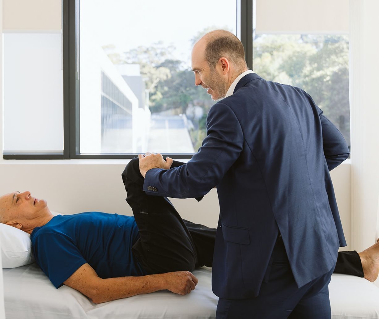
How Do Labral Tears Impact Your Health?
- Asymptomatic tears: Whilst labral tears can be painful, it is important to note that labral ‘tears’ are actually also very common findings on hip MRIs performed on people without any hip symptoms at all. Some age-related changes in the MRI appearance of the labrum are normal, so most labral ‘tears’ don’t require surgery.
- Pain and Discomfort: Individuals with hip labral tears often experience pain in the hip or groin, particularly during hip movements such as deep squatting, sitting, prolonged standing, and fast walking
- Limited Range of Motion: The tear can restrict the hip joint's range of motion, making it difficult to perform certain movements, such as bending or rotating the hip.
Risk Factors for Labral Tears
- Athletes: Activities involving repetitive hip movements, such as running, jumping, or twisting, increase the risk of hip labral tears.
- Femoroacetabular Impingement (FAI): Individuals with FAI, a condition with abnormal contact between the ball and socket of the hip joint, are more prone to hip labral tears.
- Hip Dysplasia: Individuals with hip dysplasia, a condition where the hip socket is abnormally shallow, may be more prone to developing hip labral tears due to increased stress on the labrum.
- Age and Gender: While hip labral tears can occur at any age, they are more commonly diagnosed in young adults and athletes. Additionally, females may be at a higher risk than males due to differences in hip anatomy. Degenerative labral tears can be found in most people with hip arthritis.
Causes of Labral Tears
- Structural Abnormalities: Certain structural abnormalities of the hip joint, such as hip impingement or hip joint laxity, can predispose individuals to hip labral tears. Most labral tears occur without a clear injury or trauma.
- Degenerative tears: Osteoarthritis and inflammatory arthritis of the hip often lead to labral tears, where the labral tear is a small part of the overall joint degeneration.
- Traumatic Injury: A sudden, forceful impact or trauma to the hip joint, such as a fall or sports-related collision, can cause the labrum to tear. Only a small proportion of labral tears can be attributed to a clearly defined injury.
Symptoms of Labral Tears
- Hip or Groin Pain: Persistent pain in the hip joint or groin is a hallmark symptom of hip labral tears. The pain may worsen with certain movements or activities.
- Clicking or Catching Sensation: Some individuals may experience a clicking, popping, or catching sensation in the hip joint, particularly during movements such as walking or rotating the hip.
- Limited Range of Motion: A hip labral tear can cause stiffness and limited hip joint mobility, making it difficult to perform activities requiring hip movement, such as bending or squatting.
- Instability or Weakness: In some cases, individuals with hip labral tears may experience feelings of hip joint instability or weakness, particularly during weight-bearing activities.
- Asymptomatic tears: Many labral tears do not cause symptoms and are a very common finding on hip MRIs in patients without symptoms.
Preventing Labral Tears
Labral tears cannot always be prevented, particularly when joint morphology or genetics are involved. However, many practical steps can significantly reduce the risk of developing a labral tear or prevent an existing tear from worsening. Prevention focuses on protecting hip mechanics, maintaining strength and flexibility, and avoiding unnecessary joint stress.
- Maintain good hip strength: Strong muscles around the hip, pelvis, and core help stabilise the joint and reduce strain on the labrum. In particular, the gluteal muscles play a key role in controlling hip movement during walking, running, and sports. An experienced physiotherapist can help with this.
- Improve movement control: Poor movement patterns, such as excessive inward knee collapse or uncontrolled hip rotation, increase stress on the labrum. Learning proper technique for lifting, running, squatting, and sport-specific movements can protect the hip joint.
- Stay flexible without forcing range: Gentle stretching helps maintain normal hip mobility. However, forcing extreme ranges of motion, especially deep hip flexion or rotation, can increase labral stress and worsen symptoms.
- Gradually increase activity levels: Sudden increases in training intensity, duration, or frequency raise the risk of hip injuries. Gradual progression allows the hip joint and surrounding muscles to adapt safely.
- Avoid repetitive overload: Repeated high-impact or rotational activities without adequate recovery can irritate the labrum. Cross-training and rest days help reduce cumulative joint stress.
- Address hip pain early: Persistent hip or groin pain should not be ignored. Early assessment enables identification of underlying issues, such as impingement or muscle imbalance, before a labral tear worsens.
- Manage hip impingement proactively: People diagnosed with femoroacetabular impingement benefit from targeted physiotherapy and activity modification. Early management can reduce repetitive labral trauma.
- Use proper footwear and surfaces: Supportive footwear and appropriate training surfaces help reduce abnormal forces transmitted through the hip during activity.
- Maintain a healthy body weight: Excess weight increases joint load and can accelerate hip stress, particularly during weight-bearing activities.
- Optimise posture and daily habits: Prolonged sitting with poor posture can maintain hip flexion, increasing labral stress. Regular movement breaks and ergonomic adjustments support joint health. Sit-stand desks can also be beneficial for people with labral-related hip pain.
- Rehabilitate fully after injury: Returning to sport or activity before strength and control are restored increases the risk of labral injury. Structured rehabilitation reduces recurrence.
Labral Tear Terminology
- Acetabular Labral Tear: This type of tear occurs within the labrum and is typically caused by trauma, repetitive movements, or underlying hip conditions.
- Paralabral Cyst: Sometimes, a hip labral tear can lead to the formation of a paralabral cyst, which is a fluid-filled sac that develops adjacent to the tear.
- Labral Detachment: In severe cases, the labrum may become completely detached from the acetabulum, resulting in significant instability and functional impairment of the hip joint.
- Chondrolabral junction: This is the join between the labrum and the cartilage of the inside of the socket. It is often one of the first sites of injury.
- Location of the tear: Tears may be described as anterior (at the front), anterosuperior (front + upper) or posterior (at the back). Tear location is also often described using the clock face, where 12 o’clock is directly superior and lateral on the acetabulum, 3 o’clock is anterior, and 9 o’clock is posterior, regardless of whether it is a left or right hip.
Diagnosis of Labral Tears
- Medical History and Physical Examination: Dr Williams typically begins by obtaining a thorough medical history and performing a physical examination to evaluate hip pain, range of motion, and signs of joint instability.
- Imaging Studies:
- Magnetic Resonance Imaging (MRI) is the most commonly used imaging modality for diagnosing hip labral tears. It provides detailed images of the hip joint and surrounding structures.
- Magnetic Resonance Arthrography (MRA): MRA involves injecting a contrast agent into the hip joint before MRI, thereby enhancing visualisation of the labrum and aiding detection of tears.
- X-ray: While plain X-rays often do not show the labral tear itself, they are very important in helping Dr Williams understand the underlying anatomy that may have predisposed a patient to the labral tear in the first place, such as hip dysplasia or femoroacetabular impingement.
- Diagnostic Hip Arthroscopy: A diagnostic hip arthroscopy may be performed when the diagnosis is uncertain or further evaluation is needed. During this minimally invasive procedure, a small camera is inserted into the hip joint to visualise the labrum and assess for tears or other abnormalities. Most commonly, however, a hip arthroscopy is performed only after the diagnosis is confirmed to treat a labral tear.
Treatment Options for Labral Tears
Conservative Treatment Options
Most labral tears can be managed without surgery.
- Rest and Activity Modification: Resting the hip joint and avoiding activities that exacerbate symptoms can help reduce pain and inflammation associated with hip labral tears.
- Physical Therapy: A structured physical therapy program focused on hip-strengthening, flexibility, and stability exercises can improve hip joint function and alleviate symptoms.
- Non-Steroidal Anti-Inflammatory Drugs (NSAIDs): Over-the-counter or prescription NSAIDs may be recommended to help reduce pain and inflammation associated with hip labral tears.
- Corticosteroid Injections: In some cases, corticosteroid injections may be used to provide temporary relief of hip pain and inflammation associated with labral tears. This can allow the body time to heal and the patient to engage in joint rehabilitation to reduce symptoms.
Surgical Treatment Options
- Hip Arthroscopy: Arthroscopic surgery is the most common surgical approach for treating hip labral tears. During this minimally invasive procedure, the torn portion of the labrum is repaired or removed, and any underlying hip conditions contributing to the tear, such as FAI, may be addressed.
- Labral Reconstruction: In cases of severe or irreparable labral tears, labral reconstruction may be necessary. This involves using donor tissue or tendon autograft (such as a portion of the patient’s own iliotibial band) to reconstruct the damaged labrum.
- Hip Preservation Surgery: For younger individuals or athletes with hip labral tears and structural hip abnormalities, other typed of hip preservation surgery may be performed to realign the hip joint and prevent further damage. Different surgical techniques such as periacetabular osteotomy, femoral derotation osteotomy or surgical hip dislocation may be required.
- Hip replacement surgery: In older patients (over 55) with labral tears and associated early arthritic changes, hip replacement or resurfacing may be a more reliable alternative to hip arthroscopy with regard to long-term pain relief.
What if Labral Tears are Untreated?
- Often, not much: The term labral ‘tear’ carries the connotation that an acute injury has occurred, and often a sense of urgency that it needs to be repaired. Given that most labral changes reported as ‘tears’ happen gradually over time, and are very common in asymptomatic people (those with no pain), not all labral tears need active treatment. By the time patients book to see Dr Williams for assessment, however, they have often been managing the pain for several months without improvement and have tried physiotherapy and pain relief without success. These patients may benefit from targeted non-operative or operative treatment.
- Degenerative Changes: Untreated hip labral tears may gradually lead to degenerative changes in the hip joint over time, increasing the risk of osteoarthritis and joint deterioration.
Leaving painful labral tears untreated can worsen symptoms, lead to long-term joint damage, and reduce quality of life. Early detection, accurate diagnosis, and prompt initiation of appropriate treatment can help optimise outcomes and preserve hip joint function.